EMG (biofeedback) and RUSI (rehabilitative ultrasound imaging)
EMG (biofeedback)
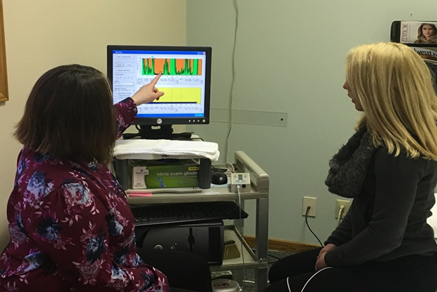
EMG (biofeedback) is completed by placing small sensors on the pelvic floor muscles (PFM) to record muscle activity during active contractions. The information collected will include your muscle resting tone, muscle endurance, and how quickly you are able to recruit muscle fibers and relax them. This assists the therapist in creating a personal exercise program to increase the strength and holding power of the muscles that control bowel and bladder. This program also may decrease muscle tension if there is high activity at rest.
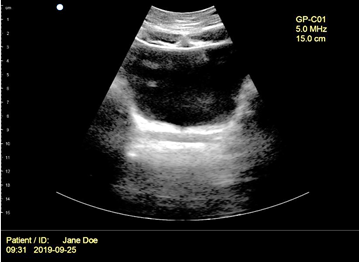
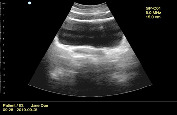
RUSI (rehabilitative ultrasound imaging)
Midwest Physical Therapy Services performs “Rehabilitative Ultrasound Imaging” (RUSI) with our patients experiencing pelvic floor dysfunction. This includes providing visual feedback to you and your physical therapist to improve clinical outcomes.
Surface EMG provides a quantitative value of the motor activity of the PFM, but cannot validate PFM lift which is associated with bowel and bladder function. Frequently, patients are bearing down with their pelvic floor when they think they are lifting it. RUSI can provide information on muscle size as well as motor activity patterns for the PFM, abdominal wall, and lumbar stabilizers. This is real time so you can actually see your muscles moving on the screen as you breathe, while attempting to contract and relax muscles, or perform specific exercises. The transducer head may be used with you lying down, sitting, standing, or with weight shifts to provide feedback of muscle use during a movement pattern.
Using RUSI, you can observe your overactive PFM and/or abdominal muscles on the screen. You will instantly receive verbal cues from the therapist and are able to see your corrected activation. This has been especially valuable for patients with constipation as they work on their home program.
Many patients contract their abdominal wall incorrectly and this is instantly visible on the screen with RUSI and easy to correct during the same session. We can assess for your correct co-contraction of the transverse abdominal muscle and PFM while you are performing lower extremity movement.
RUSI is an accurate tool to measure the amount of separation of a Diastasis Recti (DRA). We are able to determine whether the DRA is functional and stable during exercise and certain activities.